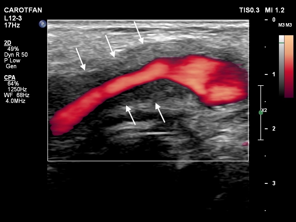
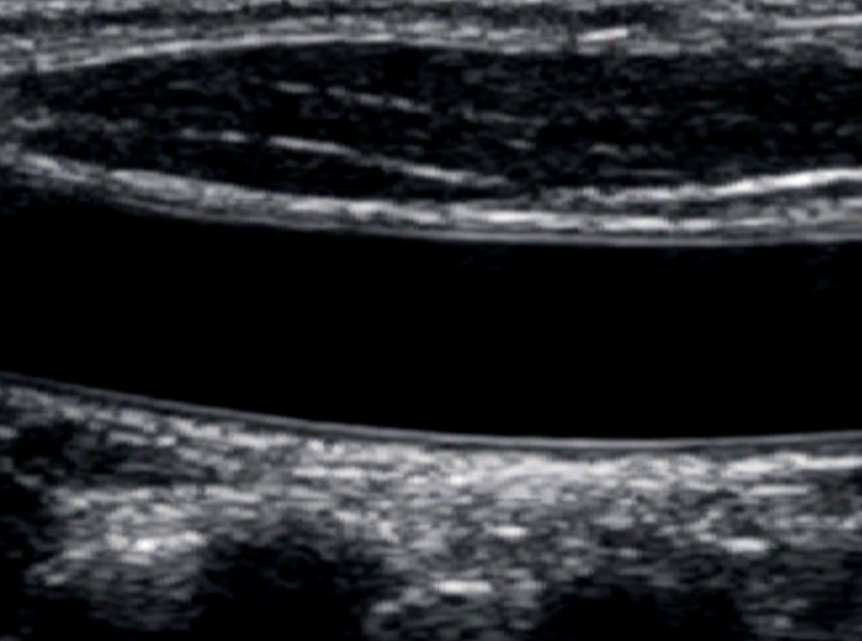
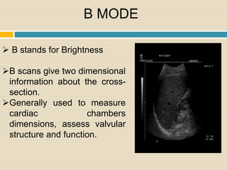
A TIPIC Ultrasonographic B-Mode Imaging of the Common Carotid
By A Mystery Man Writer
Introduction/Patient Description Extracranial carotid duplex ultrasonography (DUS) was requested within 2 weeks after sudden onset of unilateral, evolving, neck pain. Signs and symptoms related to a 53 year-old man included local swelling, skin changes, increased, local sensations, and high sensitivity to palpation. Atherosclerotic risk factors were not noted. He had contralateral radiation therapy, neck and

Colour Doppler evaluation of extracranial carotid artery in patients presenting with features of cerebrovascular disease: A clinical and radiological correlation
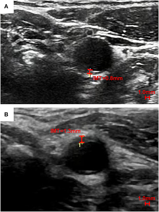
Effects of Smoking on Intima-Media Thickness of the Common Carotid Artery Using Ultrasonography, Artery Research
Atherosclerotic carotid disease (Chapter 5) - Manual of Neurosonology
Research tools Vascular Observations through Research on Technology and Exercise Lab
Frontiers The Predictive Value of Carotid Ultrasonography With Cardiovascular Risk Factors—A “SPIDER” Promoting Atherosclerosis

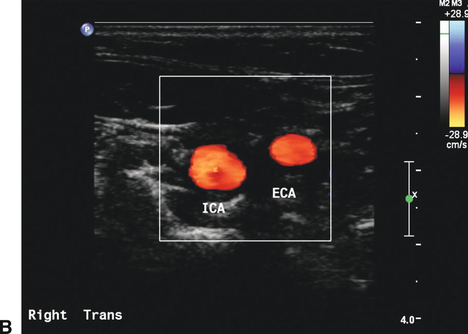
Automatic Detection of Common Carotid Artery in Transverse Mode Ultrasound Images
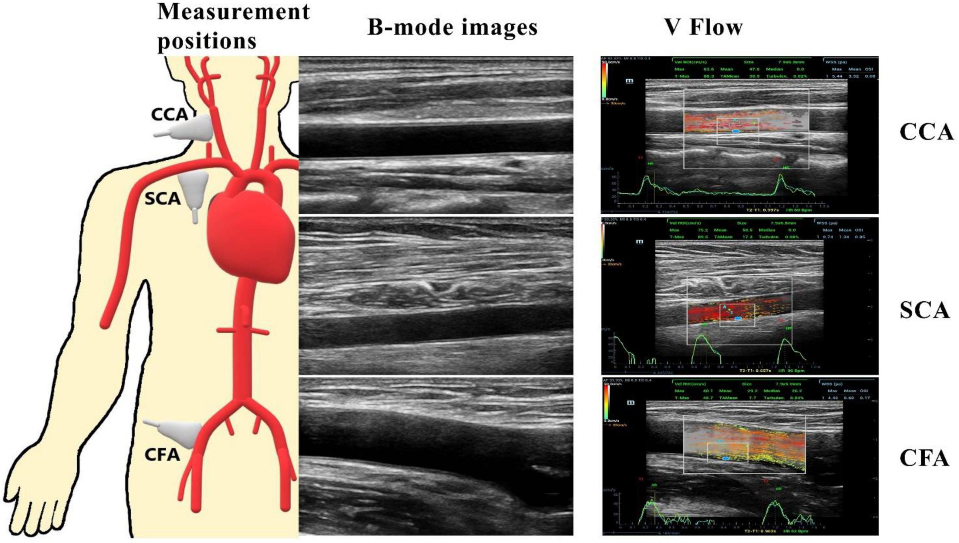
Frontiers Investigation on the differences of hemodynamics in normal common carotid, subclavian, and common femoral arteries using the vector flow technique
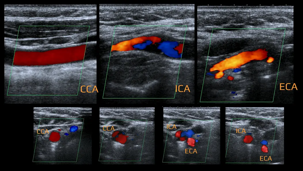
Extracranial Doppler Sonography

B-mode ultrasound image of a longitudinal section of the carotid artery
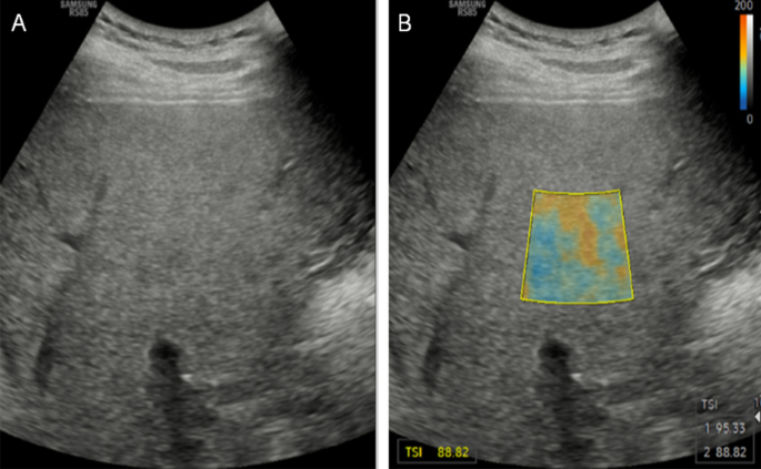
Quantitative ultrasound imaging of soft biological tissues: a primer for radiologists and medical physicists, Insights into Imaging

Manifestations of Cardiac Disease in Carotid Duplex Ultrasound Examination
Extracranial Carotid and Vertebral Arteries
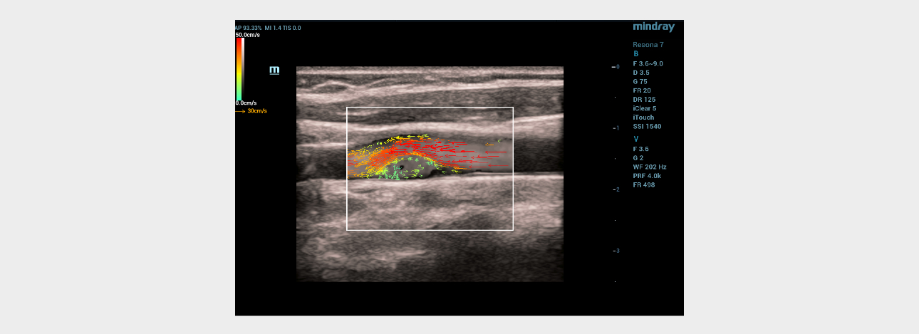
Ultrasound Journal 13 - Ultrasound Diagnostics with Carotid-Web Using V Flow Technology - Mindray
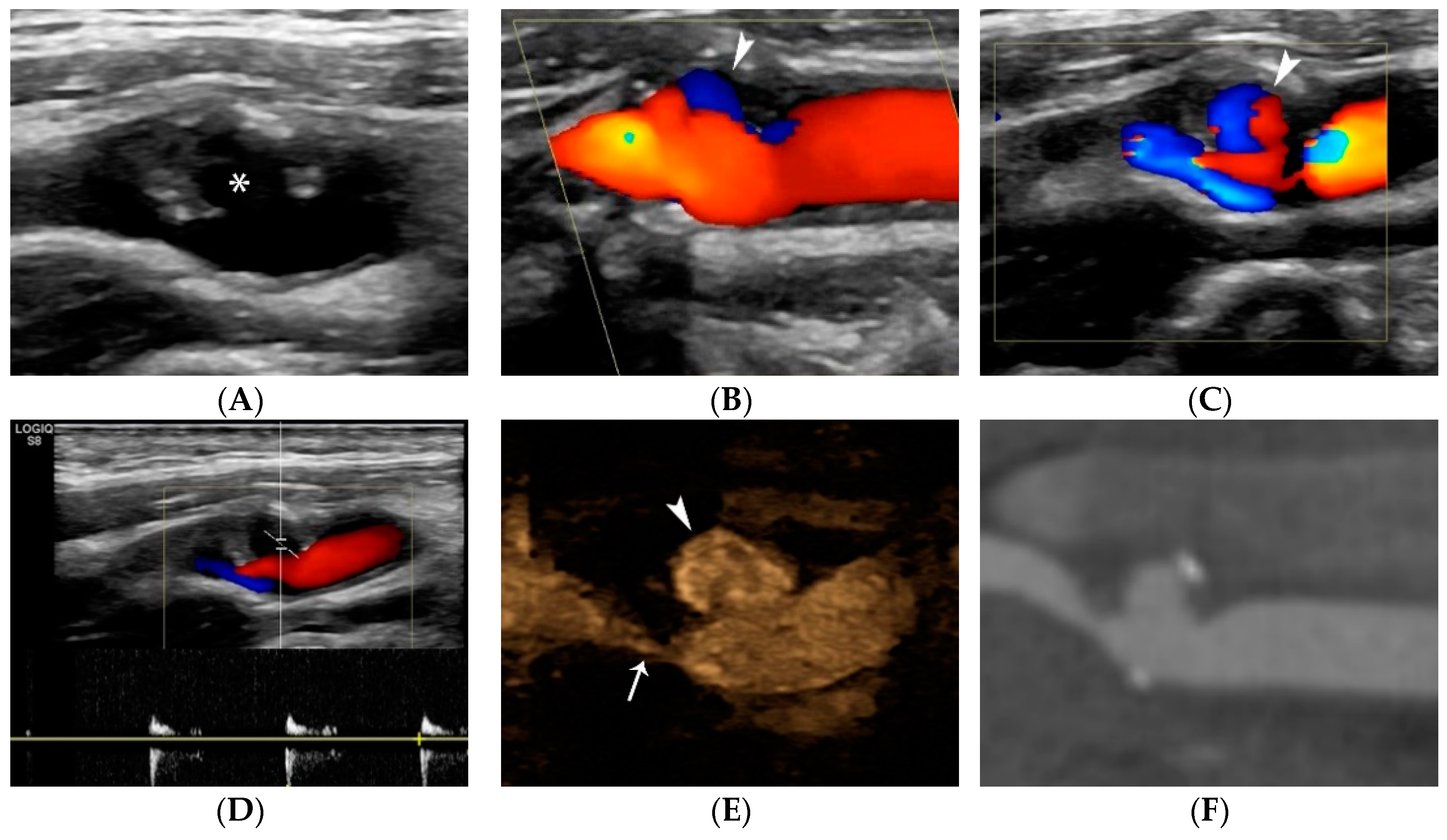
JCM, Free Full-Text
- Ultrasound imaging
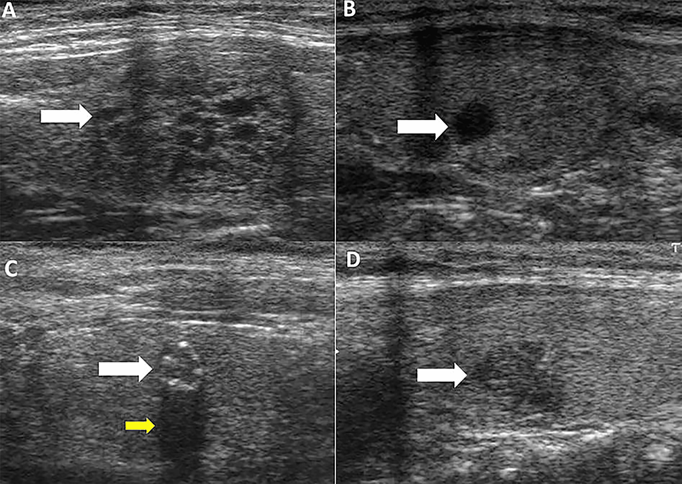
- Cureus, B-mode Ultrasound Characteristics of Thyroid Nodules With High-Benign Probability and Nodules With Risk of Malignancy
- Enhance the Range of Your LEAF

- FlightAware Band Pass Signal Filter, Dual 978-1090 MHz
- Formulation and characterisation of drug-loaded antibubbles for image-guided and ultrasound-triggered drug delivery - ScienceDirect