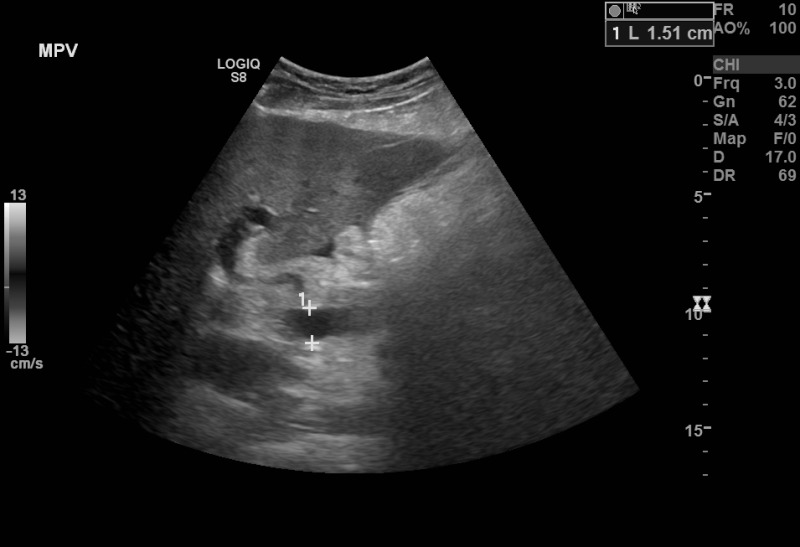
Figure, B-Mode ultrasound showing main portal] - StatPearls - NCBI Bookshelf
By A Mystery Man Writer
B-Mode ultrasound showing main portal vein diameter of 15.1 millimeters. This is an indirect finding of portal hypertension. Contributed by Brian Covello, MD
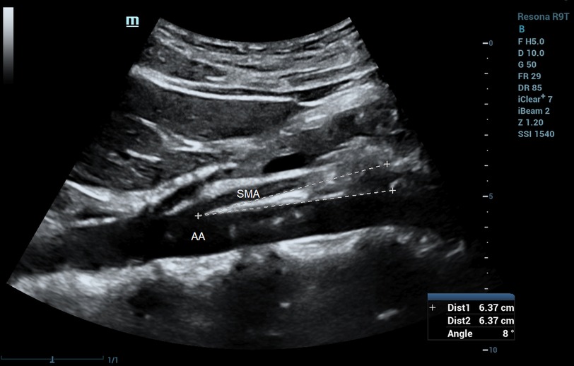
Frontiers Ultrasound characteristics of abdominal vascular compression syndromes
Can a false negative for a carotid ultrasound be the result of incorrect technique by a technician? - Quora

cardio, Department of Emergency Medicine
Salivary gland ultrasound in primary Sjögren's syndrome

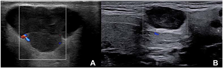
Sonography of a Typical Parathyroid Adenoma: Solitary Parathyroids as Seen on Ultrasound

Clinical Practice and Cases in Emergency Medicine Volume 4 Issue 3 by Western Journal of Emergency Medicine - Issuu

Gallbladder hydrops, Radiology Case

Estimating Ejection Fraction in a Patient with Acute-On-Chronic Systolic Heart Failure - POCUS 101
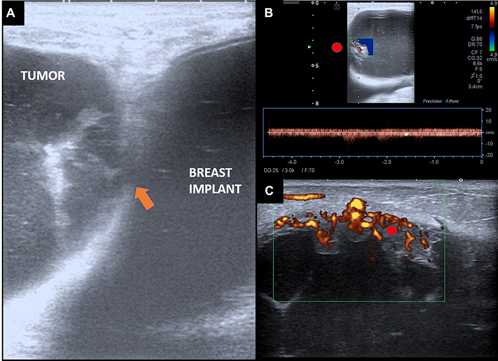
The Breast Tumor Microenvironment: Could Silicone Breast Implant Elici

Chapter 3 - Third Trimester Measurements, PDF, Pregnancy

An image classification deep-learning algorithm for shrapnel detection from ultrasound images

Frontiers Ultrasound characteristics of abdominal vascular compression syndromes

Links To And Excerpts From Comprehensive Assessment of Fluid Status by Point-of-Care Ultrasonography With Additional Resources On The Topic - Tom Wade MD