Ilium, Radiology Reference Article

By A Mystery Man Writer
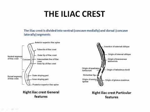
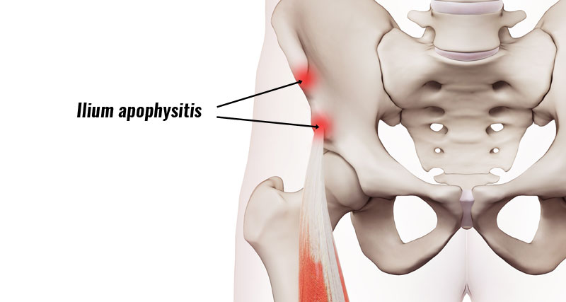
The ilium (plural: ilia) is one of the three bony components of the innominate bone: ilium, ischium, and pubis. These are individual bones in the immature skeleton which fuse to form one bone

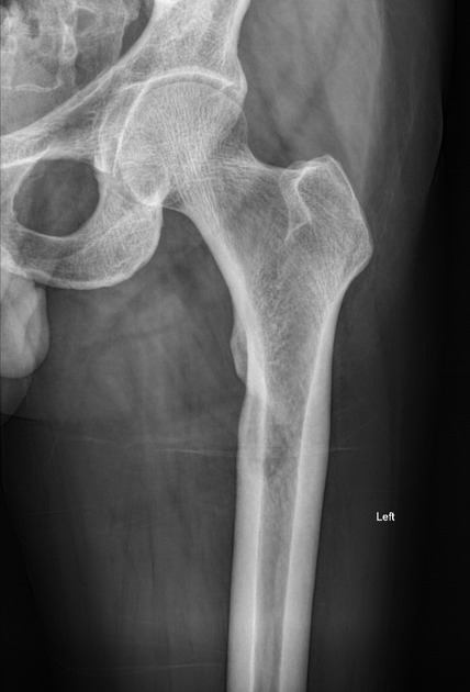
X ray pelvis anteroposterior view on the day of trauma showing

Iliac hyperdense line: a new radiographic sign of gluteal muscle contracture

Society of Skeletal Radiology– white paper. Guidelines for the diagnostic management of incidental solitary bone lesions on CT and MRI in adults: bone reporting and data system (Bone-RADS)
A) X-ray of pelvic bone showing bone destruction and irregular
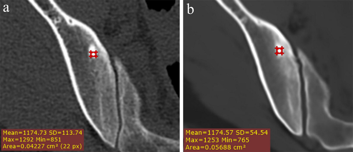
AP radiograph of the pelvis (a) and axial CT (b) in a 57-year-old

Pelvis series, Radiology Reference Article
MR Imaging of the Pelvic Bones: The Current and Cutting-Edge Techniques - Journal of the Belgian Society of Radiology

An axial CT cut of the left iliac wing at the level of the ASIS and

Radiographic examination of the horse - vet-Anatomy

MRI images showing lesion in right ilium bone.

Materials, Free Full-Text
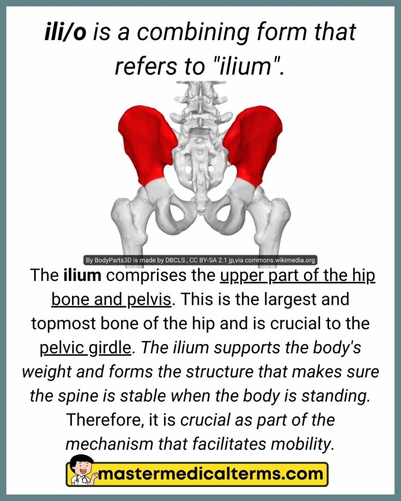
ili/o - Master Medical Terms
A) Pelvic orthography: The bone density of ilium and femur increased
Osteolytic bone lesion, Radiology Reference Article

Computed tomography shows erosions of the right ilium, involving the SI
- Under Armour Essential Fleece Cargo Joggers

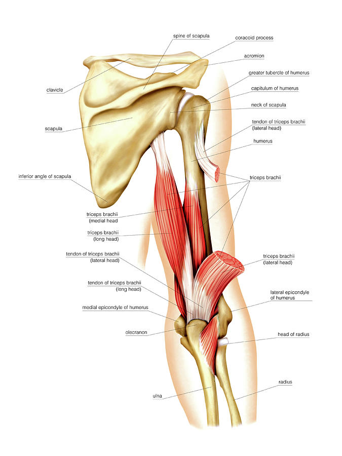
- Back Arm Muscles Photograph by Asklepios Medical Atlas - Fine Art
- eczipvz T Shirts for Men Graphic Men's Graphic Print Golf Polo Shirt Short Sleeve Lightweight Lapel T-Shirt Cotton Shirts, Yellow, X-Large : : Clothing, Shoes & Accessories

- L. Chlorine Free Organic Cotton Ultra Thin Liners Regular Absorbency, 100 count - Kroger
- Pastel Blue High-waisted Thong - Stretch Lace Underwear