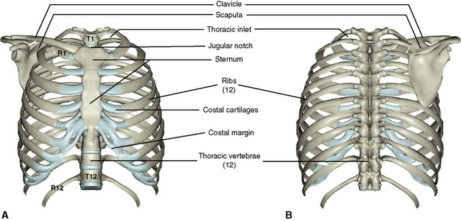
Figure 3 from Relevant surgical anatomy of the chest wall.

By A Mystery Man Writer
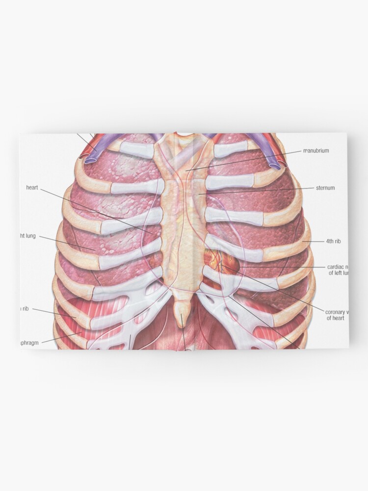
Fig. 3. Anterior chest wall showing the sternum. Note where the costal cartilages articulate with the sternum. In the intercostal space lie different structures: several kinds of intercostal muscles, intercostal arteries and associated veins, lymphatics, and nerves. (From Rendina EA, Ciccone AM. The intercostal space. Thorac Surg Clin 2007;17(4):491e501; with permission.) - "Relevant surgical anatomy of the chest wall."
3: The Thorax Pocket Dentistry
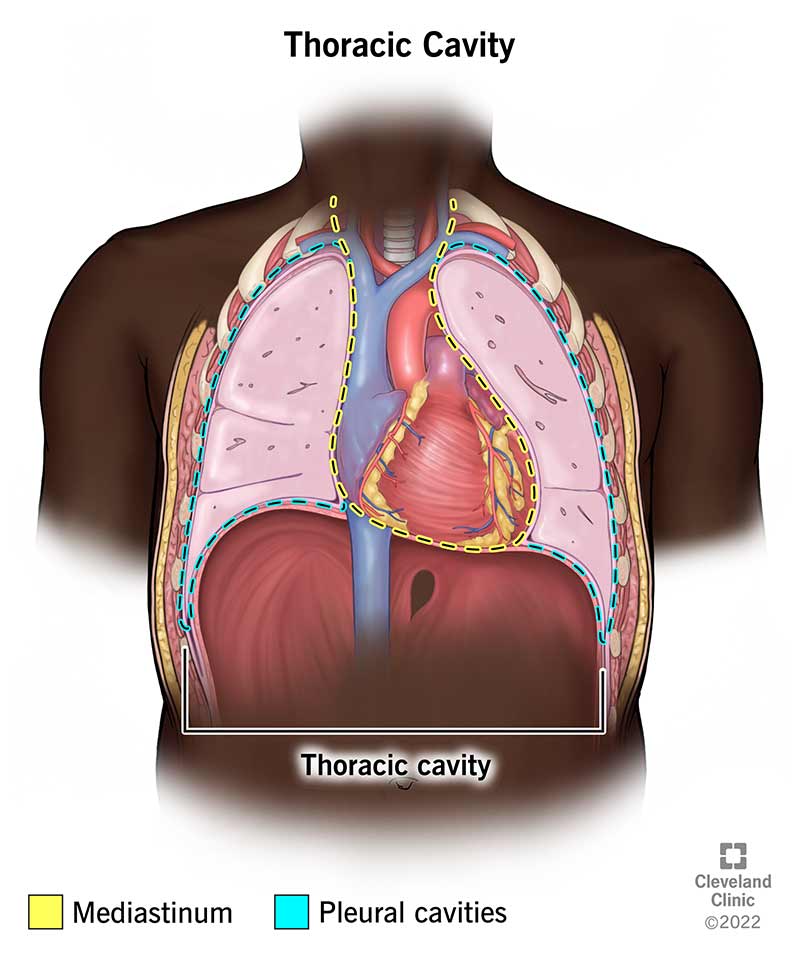
Thorax: Anatomy, wall, cavity, organs & neurovasculature
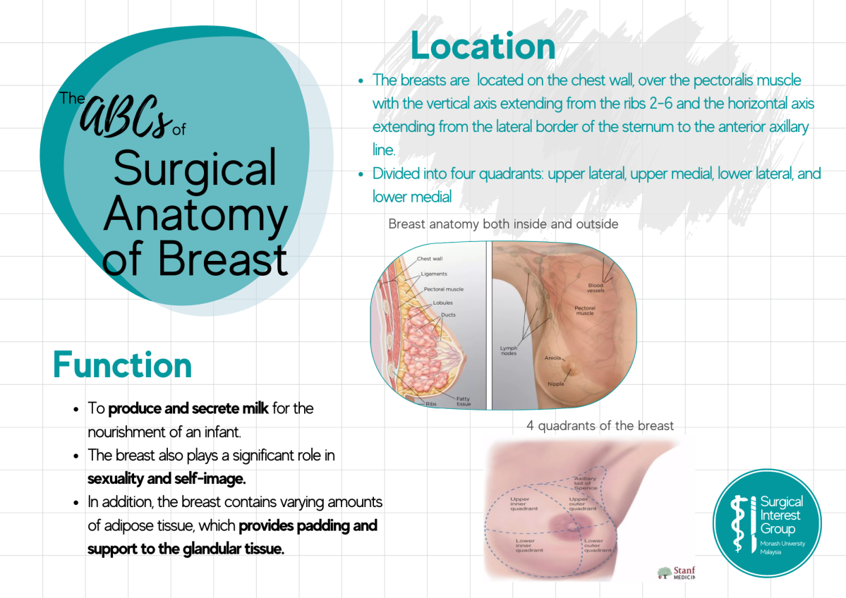
ABCs of: Surgical Anatomy of the Breast – Surgical Interest Group of Monash University Malaysia

Breast anatomy: Functions and how to check for breast cancer
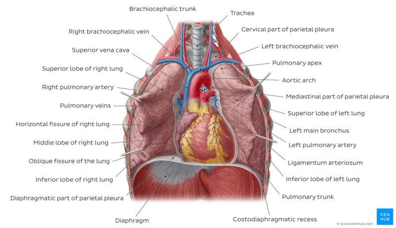
Lung Anatomy - Physiopedia

Minimally Invasive Surgical Correction of Chest Wall Deformities in Children (Nuss Procedure) - Advances in Pediatrics

Relevant anatomy for the Pecs blocks : nerves and muscles (right

Musculoskeletal Imaging of Chest Wall Injuries in Athletes - ARRS InPractice

Disorders of the Chest Wall - TeachMeSurgery

- Jxstar Girls Metallic Leggings with Skirt Kids Shiny Sparkle Stretch Pants

- MD Seamless Maternity Panties Over Bump Mid-Thigh Kuwait

- WooBilly® Perfect fit Soft Breathable Adjustable Stretch Sexy Lace Bod - Woobilly

- Mamlyn Mesh Laundry Bag for Delicates, Wash Bag for Underwear and Lingerie, (3 Medium, 3 Small)

- Candy Cane Stripe Family Pajamas - & PET BANDANA!
