Semiquantitative (SQ) visual grading scheme for vertebral

By A Mystery Man Writer
Download scientific diagram | Semiquantitative (SQ) visual grading scheme for vertebral fractures. Genant ’ s grading scheme for a semiquantitative evaluation of vertebral fracture. The drawings illustrate normal vertebrae (top row) and mild to severe fractures (respectively in the following rows). The size of the reduction in the anterior, middle, or posterior height is reflected in a corresponding to fracture grade, from 1 (mild) to 3 (severe) from publication: Vertebral morphometry: Current methods and recent advances | Vertebral fractures are the hallmark of osteoporosis and are associated with increased morbility and mortality. Because a majority of vertebral fractures often occur in absence of specific trauma and are asymptomatic, their identification is radiographic. The two most widely | Vertebrates, Spinal Fractures and Osteoporosis | ResearchGate, the professional network for scientists.

/cms/attachment/cf3

Infrared Fiber Optic Probe Evaluation of Degenerative Cartilage Correlates to Histological Grading - Arash Hanifi, Xiaohong Bi, Xu Yang, Beril Kavukcuoglu, Ping Chang Lin, Edward DiCarlo, Richard G. Spencer, Mathias P.G. Bostrom

Daniele DIACINTI, Sapienza University of Rome, Rome, la sapienza, Department of Radiological, Oncological and Pathological Sciences

EPOS™
PPT - Vertebral shape: automatic measurement by DXA using overlapping statistical models of appearance PowerPoint Presentation - ID:468306
VIBe (Validated Intraoperative Bleeding) Scale

PDF) Vertebral morphometry: Current methods and recent advances

Cornelis KUIJK, Chair Department of Radiology and Nuclear Medicine, MD PhD (Prof Dr), Amsterdam University Medical Center, Amsterdam, VUmc, Department of Radiology

Advanced trends in magnetic resonance imaging in assessment of lumbar intervertebral degenerative disk disease, Egyptian Journal of Radiology and Nuclear Medicine
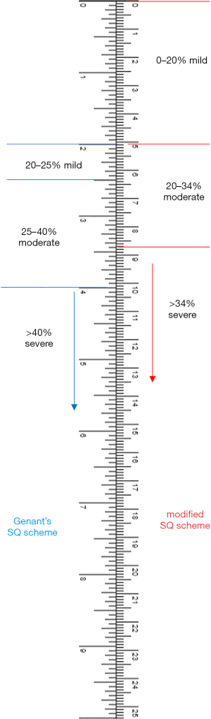
A modified semi-quantitative (mSQ) grading scheme for osteoporotic vertebral fracture in elderly women - Wáng - Quantitative Imaging in Medicine and Surgery
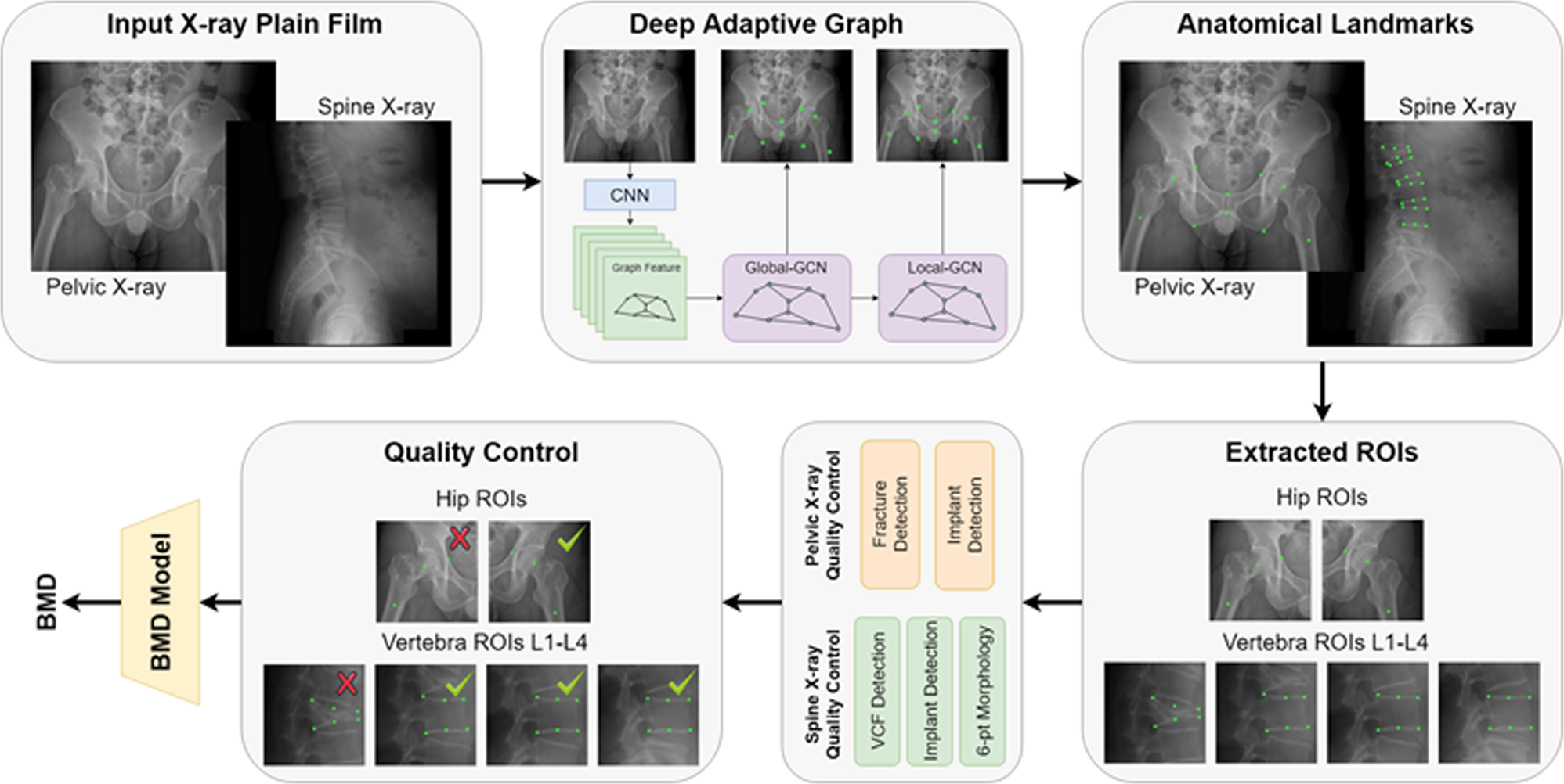
Automated bone mineral density prediction and fracture risk assessment using plain radiographs via deep learning

Cornelis KUIJK, Chair Department of Radiology and Nuclear Medicine, MD PhD (Prof Dr), Amsterdam University Medical Center, Amsterdam, VUmc, Department of Radiology

Vertebral body fractures of unknown origin in cancer patients receiving MDCT: reporting by radiologists and awareness by clinicians, SpringerPlus
- Medical Grade Compression Socks 20-30 mmHg - My Ice Wrap
- 7 Reasons to Buy/Not to Buy ZFiSt Medical Grade Sport Compression Socks
- 20-30 Compression Medical Grade Stockings for Men and Women, Knee High, Open Toe
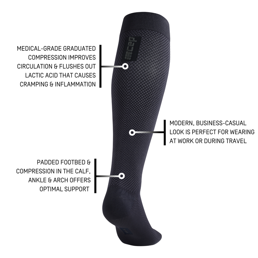
- Men's Allday Tall Compression Socks, Business Socks
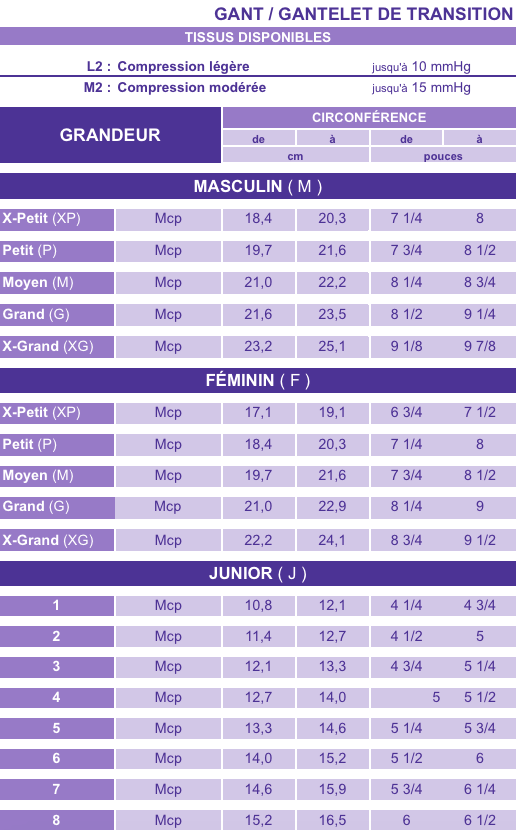
- INTERIM L2 & M2 compression glove - open tips - wrist - Medical Grade - Dermagate
- Is Disney Dreamlight Valley's Gold Edition worth it? - Dot Esports

- SKIMS review: I tried (almost) everything Kate Moss modeled
- Gibobby Yoga Shorts for Women Cotton Women's High Waist Biker Shorts Workout Yoga Running Gym Compression Spandex Shorts Side Pockets gjigj7 Army Green : ספורט ופעילות בחיק הטבע

- Women's Riders Lee Work Pants Brown Slimming Tummy Control Panel

- Women's panties Tommy Hilfiger Thong 1P - hawaiian coral, Tennis Zone