Thursday, Oct 03 2024
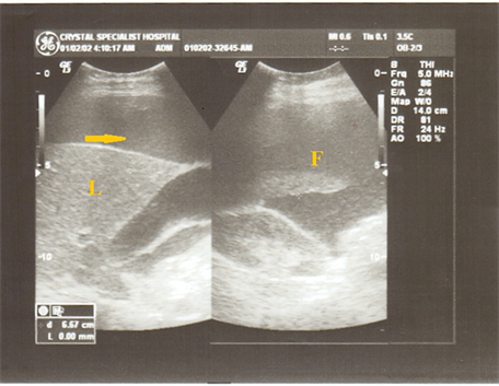
Ultrasound: Small pocket of ascites.

By A Mystery Man Writer

Figure 16 from Ultrasound for Detection of Ascites and for

Ascites, Radiology Reference Article

Figure 20 from Ultrasound for Detection of Ascites and for

– Emergency Medicine EducationUnlocking Common ED
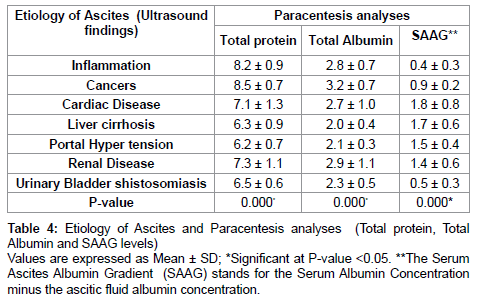
Evaluation of Ascites and its Etiology Using Ultrasonography

Troubleshooting Paracentesis Using POCUS – POCUS Journal

Ascites Fluid - an overview
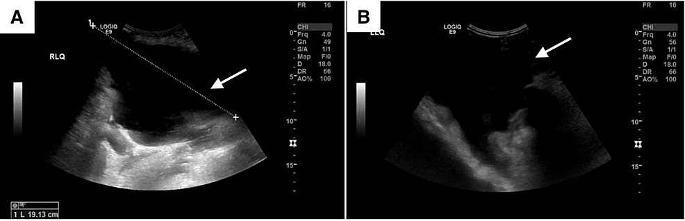
Comparative Ultrasound Review of Free Intra-Peritoneal Fluid (Ascites)

Figure 1 from Comparative Ultrasound Review of Free Intra

Figure 9 from Ultrasound for Detection of Ascites and for Guidance

Missing Inferior Vena Cava on POCUS: A Case of Left-Sided IVC with
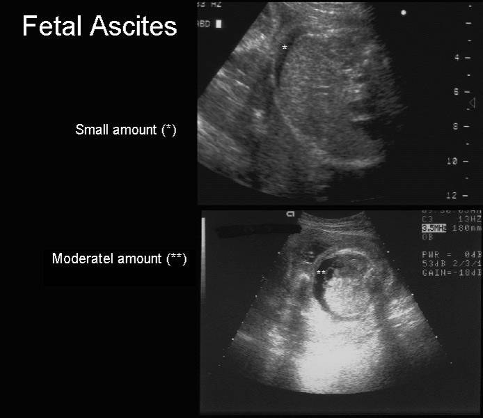
Ultrasound of Fetal Ascites

Best Practices: Paracentesis
Cureus Abdominal Wall Hematoma Secondary to Dissection of the
Related searches
- Little Baby Pocket bietet exklusive Babyprodukte

- The 17 Best Small Pocket Knives in 2024, Ranked!

- Rabbit R1: The cute little pocket-size viral AI device that can do everything for you - India Today

- Polly Pocket Tiny Pocket Places Lila Pet Compact with Doll

- Polly Pocket Tiny Pocket World, Lila – Square Imports

Related searches
- Zikra Bra And Panty in Mustafabad,Delhi - Best Lingerie Retailers in Delhi - Justdial

- Women's Maidenform 40760 Classics Microfiber and Lace Boyshort Panty (Lavender Picnic Ditsy 6)

- Adidas high waisted short leggings , Women's Fashion, Activewear

- Bookstore

- Sparkly Bikini Top - Bralette Bikini Top - Lurex Knit Bikini Top - Lulus

©2016-2024, changhanna.com, Inc. or its affiliates