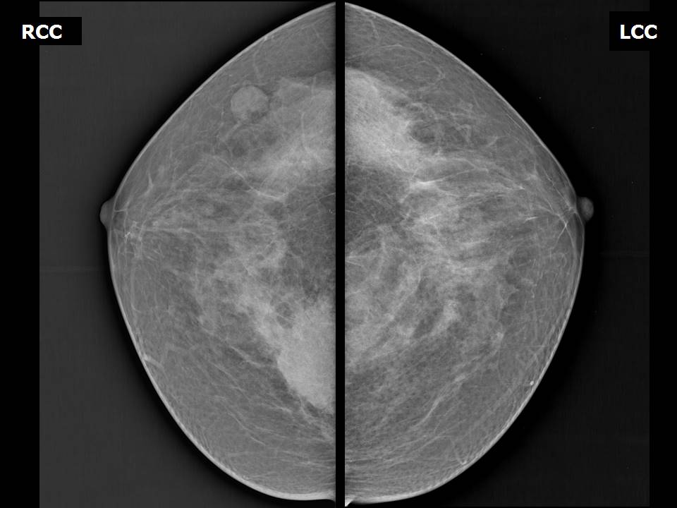
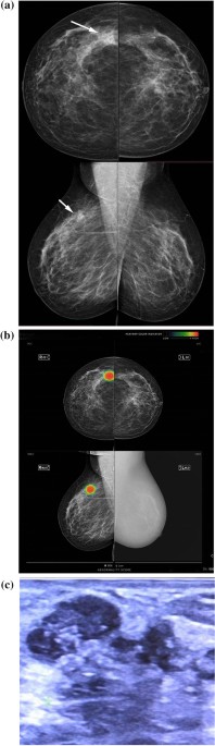
A 60-year-old patient presented by a lump in the left breast

By A Mystery Man Writer
Download scientific diagram | A 60-year-old patient presented by a lump in the left breast. Mammography revealed focal asymmetry in the left upper inner quadrant with microcalcifications (a, b). DBT showed left breast spiculated mass with microcalcifications as well as right breast retroareolar nodule with microcalcifications (c, d). CEM showed left breast heterogeneously enhancing upper inner quadrant mass lesion with spiculated margins and surrounding multiple satellite lesions as well as right breast tiny right retroareolar homogenously enhancing mass with not circumscribed irregular margins (e, f). Breast ultrasound showed left breast irregular ill-defined mass in the left upper inner quadrant as well as right retroareolar small irregular ill-defined mass (g, h). The final diagnosis was bilateral invasive duct carcinoma from publication: Comparative study between contrast-enhanced mammography, tomosynthesis, and breast ultrasound as complementary techniques to mammography in dense breast parenchyma | Background Mammography is accused of having low sensitivity and specificity in dense breast parenchyma. Also, women with dense breasts show an increased risk of developing breast cancer. Breast ultrasound has been used for several years for a better characterization of breast | Breast Ultrasound, Mammography and breast | ResearchGate, the professional network for scientists.

60 year old female patient with invasive carcinoma of no specific type

Comparison of diagnostic accuracy when mastocheck alone, mammography

Enlarging Breast Mass in 69-Year-Old Woman With History of Cysts- Clinical Advisor
Atlas of breast cancer early detection
Diagnostic mammogram of a 60-year-old female patient. (a) Left

60-year-old woman with adenoid cystic carcinoma in the left breast.
Mammographically detected asymmetries in the era of artificial intelligence, Egyptian Journal of Radiology and Nuclear Medicine

PDF) Comparative study between contrast-enhanced mammography, tomosynthesis, and breast ultrasound as complementary techniques to mammography in dense breast parenchyma
A 60-year-old patient presenting a right unilateral breast mass
:max_bytes(150000):strip_icc()/iStock-865314396-5a206816845b34003674922c.jpg)
Benign Breast Changes That Mimic Breast Cancer

10 Signs of Breast Cancer to Bring to Your Doc's Attention

Male Breast Cancer and Gynecomastia, Breast Cancer 101
Breast Fibroadenoma Imaging: Practice Essentials, Radiography, Magnetic Resonance Imaging
- Locally Advanced Breast Cancer (LABC) and Neoadjuvant Chemotherapy
- I hate that my left boob is noticeably bigger than my right boob.

- I doesn't bother me that your left breast is two cup sizes bigger than your right breast.
- What is capsular contracture and how can it be treated?

- A 60-year-old patient presented by a lump in the left breast.
- Blue Printed sport bra worn by Addy Hanlon (Herizen Guardiola) in Dare Me (S01E01)

- Body Justo Magic John John Feminino - John John

- Janet Leigh : r/ClassicScreenBeauties

- Baggy Jeans For Men Plus Size Loose Elastic Waist Denim Street Wide Leg Pants

- Owl Wooden Jigsaw Puzzles for Adult Wooden Jigsaw Puzzles Owl Animal Shape 155 Pcs 6.5 x 12.6 Colorful Puzzles Funny Bird Puzzles Animal Shaped
