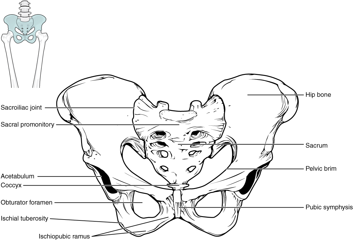
Bones of the pelvic girdle (Fig. 10.6) Diagram

By A Mystery Man Writer

Figure 10.6 Right os coxa Diagram
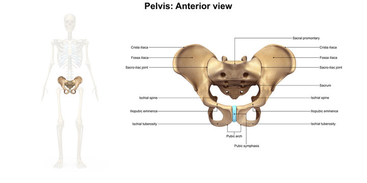
8.3 The Pelvic Girdle and Pelvis – Douglas College Human Anatomy and Physiology I (1st ed.)

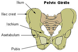
Pelvic Girdle

Pelvic girdle bones Diagram
JPM, Free Full-Text
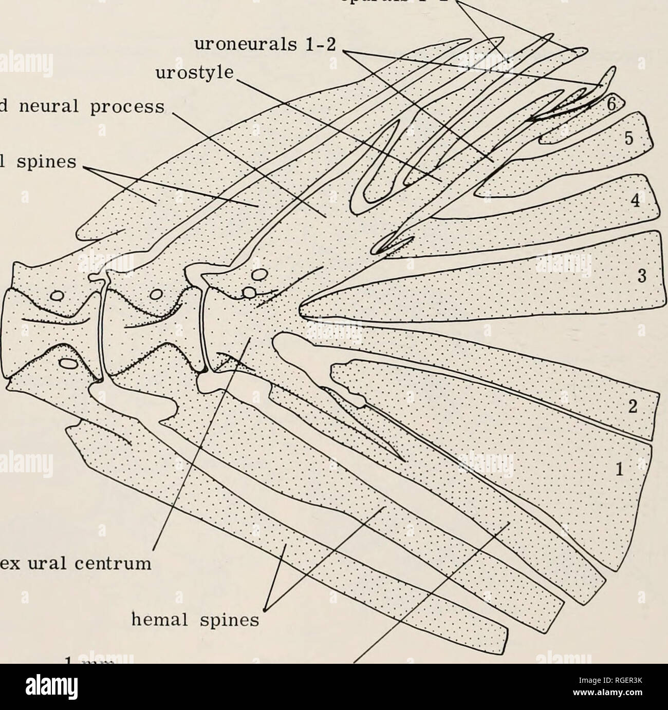
Bulletin of the Museum of Comparative Zoology at Harvard College. Zoology. pelvic bone ischiac process pelvic radials 1-4 I 1 mm I Figure 77. Saccodon wagneri (dental morph IV), 55.4 mm.

Anteroposterior radiograph of the pelvis of a 16 year-old boy with an

A) Anterior and posterior views of a symmetrical pelvis (left) and the
Elements of the comparative anatomy Elements of the comparative anatomy of vertebrates elementsofcompar00wied Year: 1886 A B FIG. 78.—PELVIC ARCH OF FROG (Rana esculenta). (A, from below; B, from the side.) II

Figure 10.6 (a) Diagram

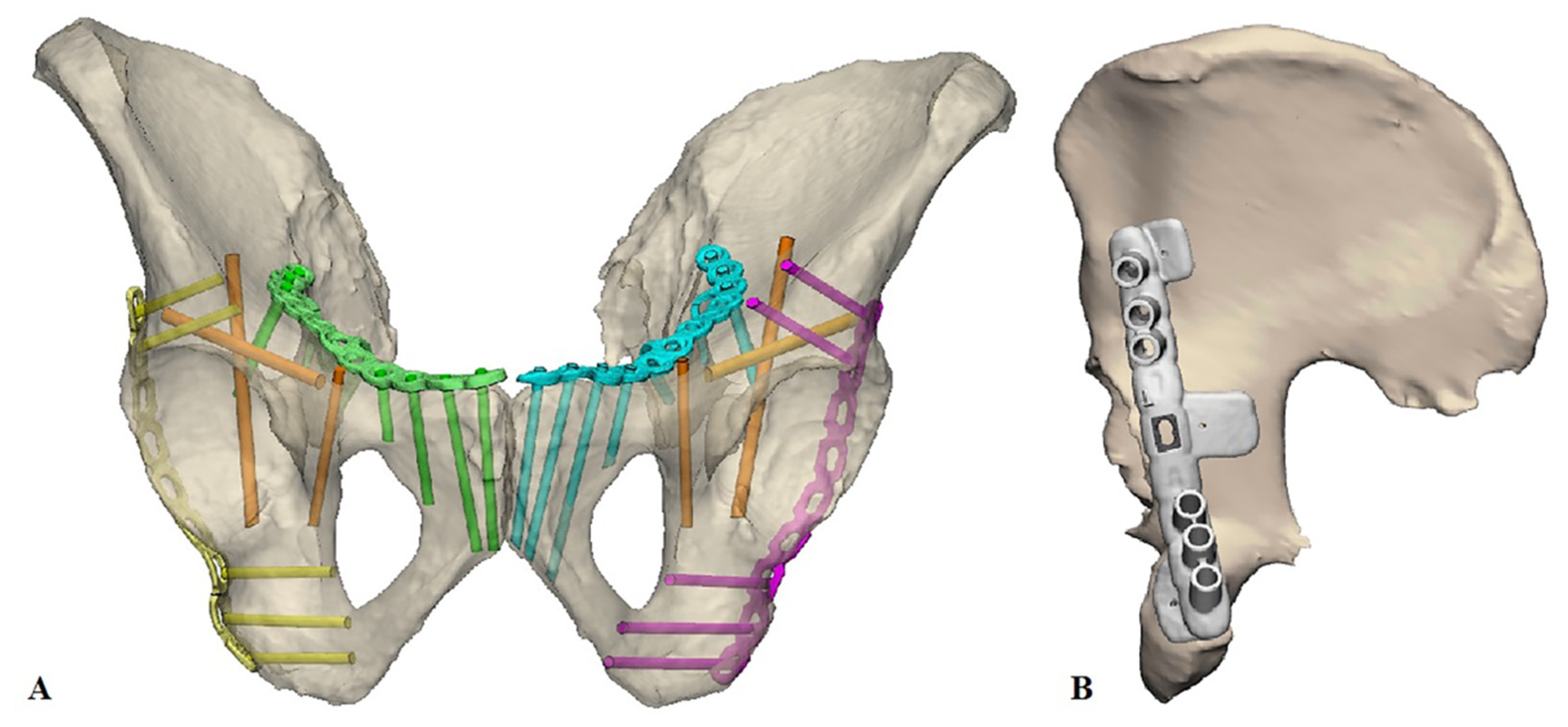
Roy-Camille classification for spinopelvic dissociation (types 1, 2
Pelvic Girdle – Coxal Bones Anatomy – Earth's Lab

Figure 10.6 Right os coxa Diagram

Pelvic mechanism of trunk straightening. (A) Muscular actions: (a)
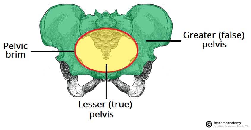
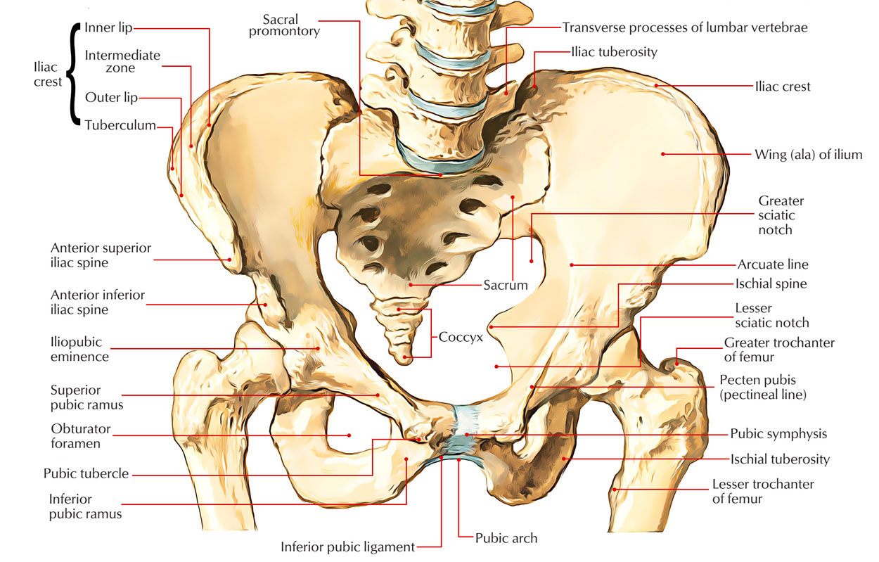
The Pelvic Girdle - Structure - Function - Assessment - TeachMeAnatomy


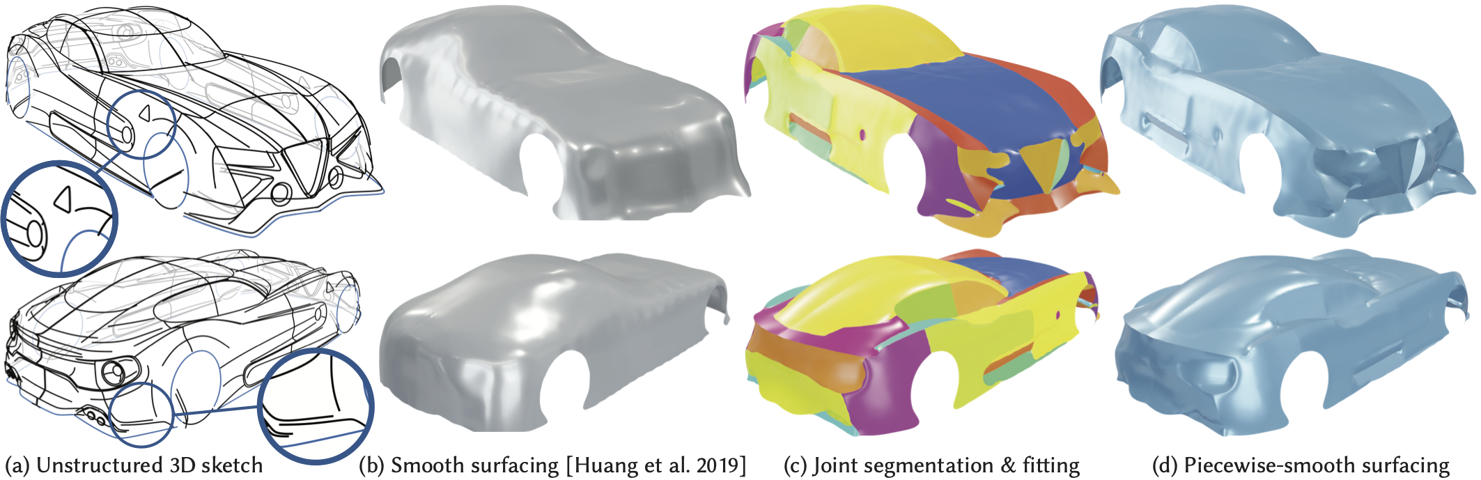
- Piecewise-Smooth Surface Fitting onto Unstructured 3D Sketches
- Pantalones casuales de color sólido para mujer, decoración de botón de tarjeta salvaje, pantalones formales, pantalones rectos en la cintura, pantalones de talla grande para mujer (rosa intenso, XL) : Ropa

- WINE BURGUNDY FLARED PALAZZO PANT SET PAIRED WITH A FLORAL PRINTED DRAPED TOP AND AN EMBROIDERED BELT. - Seasons India

- Elomi Cate Embroidered Full Cup Banded Underwire Bra (4030),40K,Alaska

- Free People, Pants & Jumpsuits
