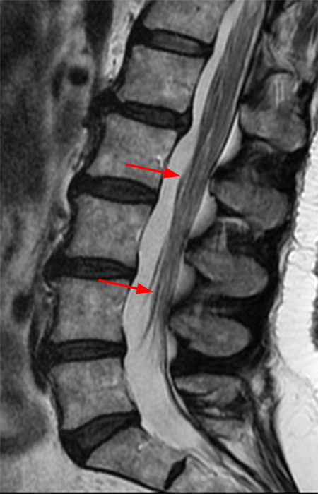
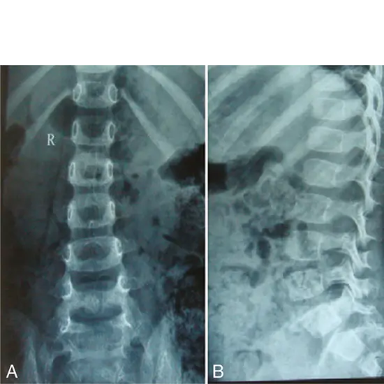
Sagittal T2 image of dorso-lumbar spine shows a hyper-intense

By A Mystery Man Writer
MR imaging of uncommon complications of transforaminal

Initial thoracolumbar spine MRI. Sagittal T2-weighted image (A

Spine SpringerLink
a & b) Coronal T2-weighted brain MRI showing a right optic nerve

Vaishali UPADHYAYA, Chief Consultant Radiologist MRI Division, Ramakrishna Mission Sevashrama, Lucknow
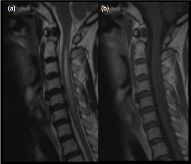
Sagittal MR FSE T1-and T2-weighted of the dorsal spine showing
Congenital and Acquired Hypertrophic Peripheral Neuropathies
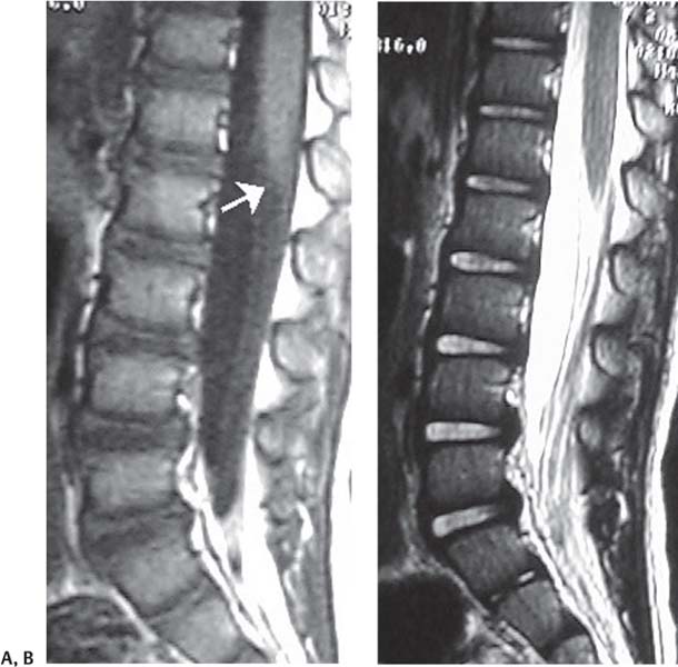
Sagittal spinal cord MRI of case 1. T2-weighted image showing a

Figure 1 from Redundant Nerve Roots of the Cauda Equina Associated

Fat in filumterminale. T2 weighted (A) and T1 weighted (B

Thoraco-lumbar spinal tumor. Sagittal T2-(A) and T1-weighted

Cureus Posterior Epidural Migration of Lumbar Intervertebral

a & b) Coronal T2-weighted brain MRI showing a right optic nerve
The Pediatric Spine


- Louisa Johnson shows off her toned tum in a frayed white crop top and high-waisted leggings

- Slim Fit Elastic Sleeveless Casual Sexy Women's Tank Top Nude Solid Color Tight Fitting Swimsuit - China Swimsuit and Sexy Jumpsuit price

- assets.myntassets.com/h_1440,q_100,w_1080/v1/asset

- 1Pcs Thick Ribbed Cuffs 40cm Soft Stretch Striped Knit Fabric Suit Down Jacket Coat Trousers Accessories
- Women's Mesh-Trimmed Michele Bralette