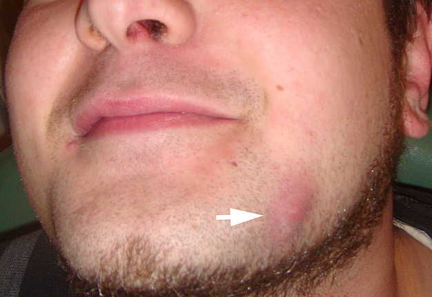
a Mandibular fistula indicated by an arrow in the apical region of dd
By A Mystery Man Writer
Download scientific diagram | a Mandibular fistula indicated by an arrow in the apical region of dd 36-37. b A fistula in the apical region of dd 46-47 (white arrows) and a red area in the mucosa (black arrows) are seen in the right lingual surface of the mandible. c Panoramic radiograph showing no bone lesions in the mandible. d Periapical x-ray with no bone involvement in the apical region of dd 46-47 from publication: Treatment of bisphosphonate-induced osteonecrosis of the jaws with Nd:YAG laser biostimulation | Osteonecrosis, Jaw and Nd:YAG Laser | ResearchGate, the professional network for scientists.

Malformations of the tooth root in humans. - Abstract - Europe PMC

a Mandibular fistula indicated by an arrow in the apical region of dd

SciELO - Brazil - Differential diagnosis and clinical management of periapical radiopaque/hyperdense jaw lesions Differential diagnosis and clinical management of periapical radiopaque/hyperdense jaw lesions

Anti-vascular endothelial growth factor antibody monotherapy causes destructive advanced periodontitis in rice rats (Oryzomys palustris) - ScienceDirect

Ultrasonographic Imaging in Periodontology

Healthcare, Free Full-Text

Satu ALALUUSUA, University of Helsinki, Helsinki, HY, Institute of Dentistry

Radiolucent lesions of the mandible: a pattern-based approach to diagnosis, Insights into Imaging
Frs hfn

Atlas of cosmetic and reconstructive periodontal surgery 3rd edition cohen by dental.id - Issuu

Dental CT: Pathologic Findings in the Teeth and Jaws

/profile/Maria-De-Souza-3/publ
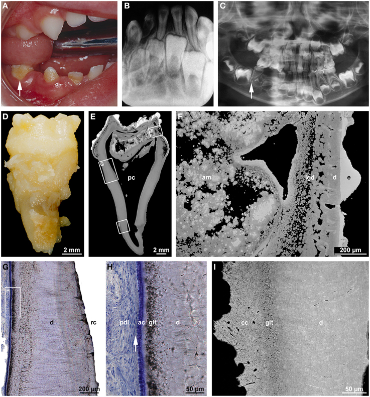
Frontiers Malformations of the tooth root in humans
Single and Multiple Odontogenic Cutaneous Sinus Tracts
- DD36 | Wolf 36 Downdraft Ventilation System - Blowers Required

- U-Joint - 1 DD x 17mm 36-Spline - Electra-Steer - 8052530
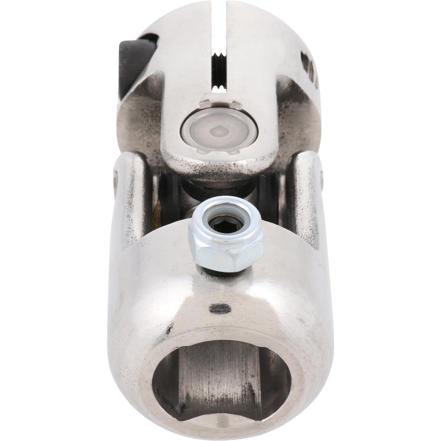
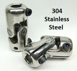
- U-Joint - 3/4 DD x 3/4 36-Spline (Ford) - Stainless Steel
- Steering Coupler - 3/4 DD x 17mm 36-Spline - Electra-Steer

- Women's Sports Bras - DD / 36 / Women's Sports Bras / Women's Bras: Clothing, Shoes & Jewelry

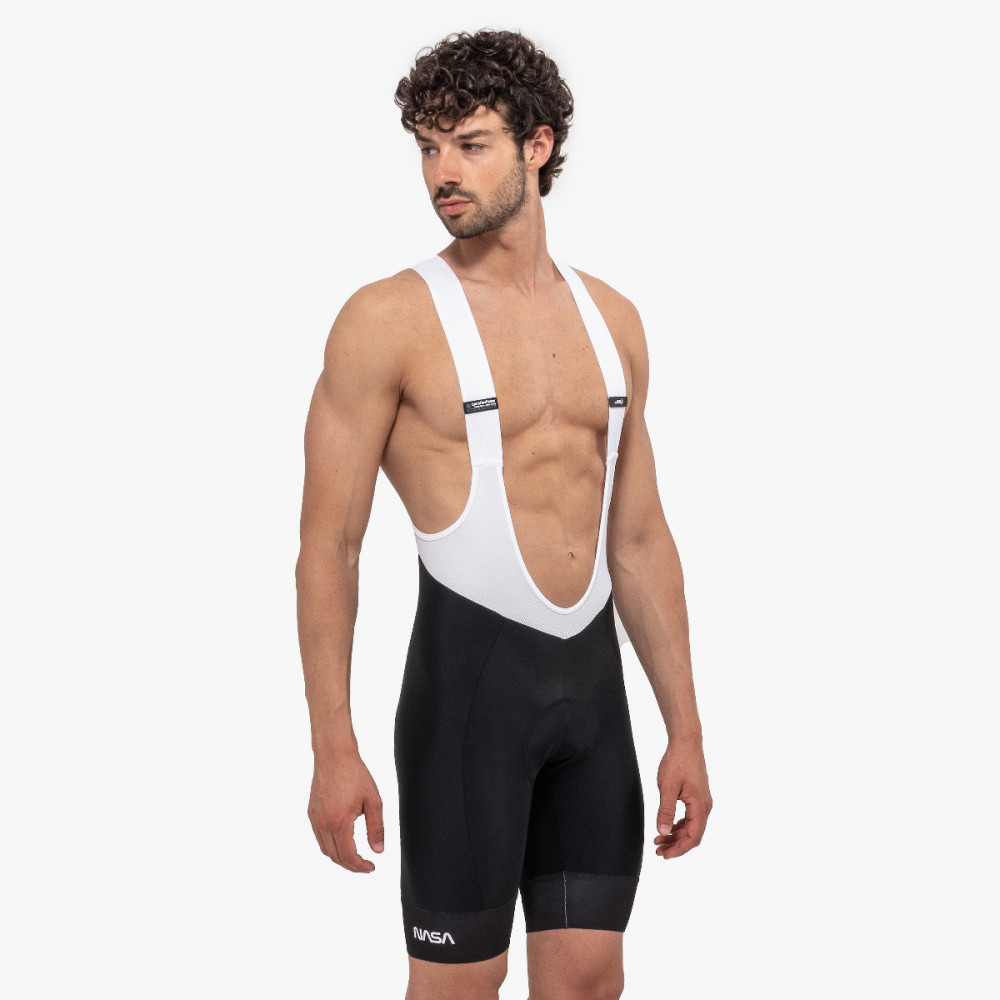
- SPACE AGENCY X-OVER CYCLING BIB SHORTS - MAN
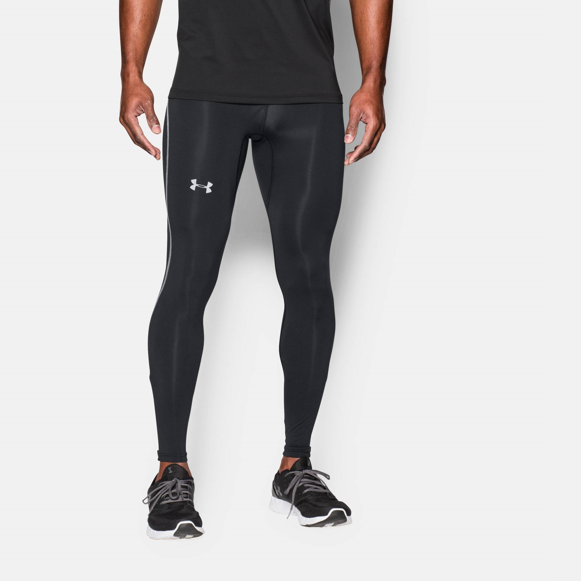
- Leggings & Tights, Under armour CoolSwitch Run. Comp. Leggin 1991
- A tendência do Jeans na moda e nos sapatos

- gvdentm Maternity Bra Women's Minimizer Bras Comfort Cushion Strap Wirefree Full Coverage Large Bust Non-Padded Br

- Seafolly Women's Standard C D Trapeze Bandeau Tankini Top Swimsuit, La Belle Black, 8 US - Discount Scrubs and Fashion