Calcification and mass abnormalities in breast mammogram scans
By A Mystery Man Writer
Download scientific diagram | Calcification and mass abnormalities in breast mammogram scans. The calcification distribution depicts tiny flecks of calcium as small white regions on the left side, while the mass is shown as a smooth, well-defined border on the right side. from publication: Multi-Graph Convolutional Neural Network for Breast Cancer Multi-Task Classification | Mammography is a popular diagnostic imaging procedure for detecting breast cancer at an early stage. Various deep learning (DL) approaches to breast cancer detection incur high costs and are prone to classify incorrectly. Therefore, they are not sufficiently reliable to | Breast Cancer, Convolution and Classification | ResearchGate, the professional network for scientists.

PDF) Multi-Graph Convolutional Neural Network for Breast Cancer Multi-Task Classification
Mammography of breast calcifications
What Do Spots on a Mammogram Mean? - Health Images

Breast Lesions Detection and Classification via YOLO-Based Fusion Models

Comparison and risk factors analysis of multiple breast cancer screening methods in the evaluation of breast non-mass-like lesions, BMC Medical Imaging

Shagufta HENNA, Lecturer
Mammography: Masses - Radiology

Brendan JENNINGS, Head of Graduate Studies
Breast calcifications mimicking pulmonary nodules
Atlas of breast cancer early detection

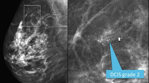
Calcifications at Digital Breast Tomosynthesis: Imaging Features and Biopsy Techniques

a) The cropping breast profile image of mdb111 for left MLO
- Does Breast Size Affect Your Risk of Breast Cancer?
:max_bytes(150000):strip_icc()/GettyImages-917730122-5af4921f3418c60038771575.jpg)
- Will a Mammogram Work for Me? FAQs about Small Breasts, Dense Breasts, and Implants, Huntsman Cancer Institute
- What are dense breasts, and why is it important for women to know if they have them?, MUSC
- Mammogram what you need to know

- Why the way microcalcifications on mammograms are regarded could
- DKNY WOMENS LITEWEAR Low Rise Mesh Trim Thong,Vanilla,Small £17.04
- Kauai teacher heading to USA Yoga national championships - The Garden Island
- Curly Braces Punctuation Mark Black Icon. Vintage Elegant Brackets Symbol For Typing Or Typography. Ornament And Vector Eps Isolated Design Element Concept For Messages And Quotes Royalty Free SVG, Cliparts, Vectors, and

- Sticky Bra Invisible Strapless Adhesive - Push up Bra for Big Busted

- Ask the Experts: What could a Weak Urine Flow be telling me about my Health? - Urology Care Foundation