Breast volumes of human subjects in three scanning positions.

By A Mystery Man Writer

3-D breast area and breast area difference (BAD) calculation in cm 2 on

Muriel Brackstone's research works London Health Sciences Centre, London and other places
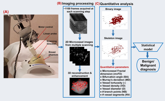
Volumetric imaging and morphometric analysis of breast tumor angiogenesis using a new contrast-free ultrasound technique: a feasibility study, Breast Cancer Research
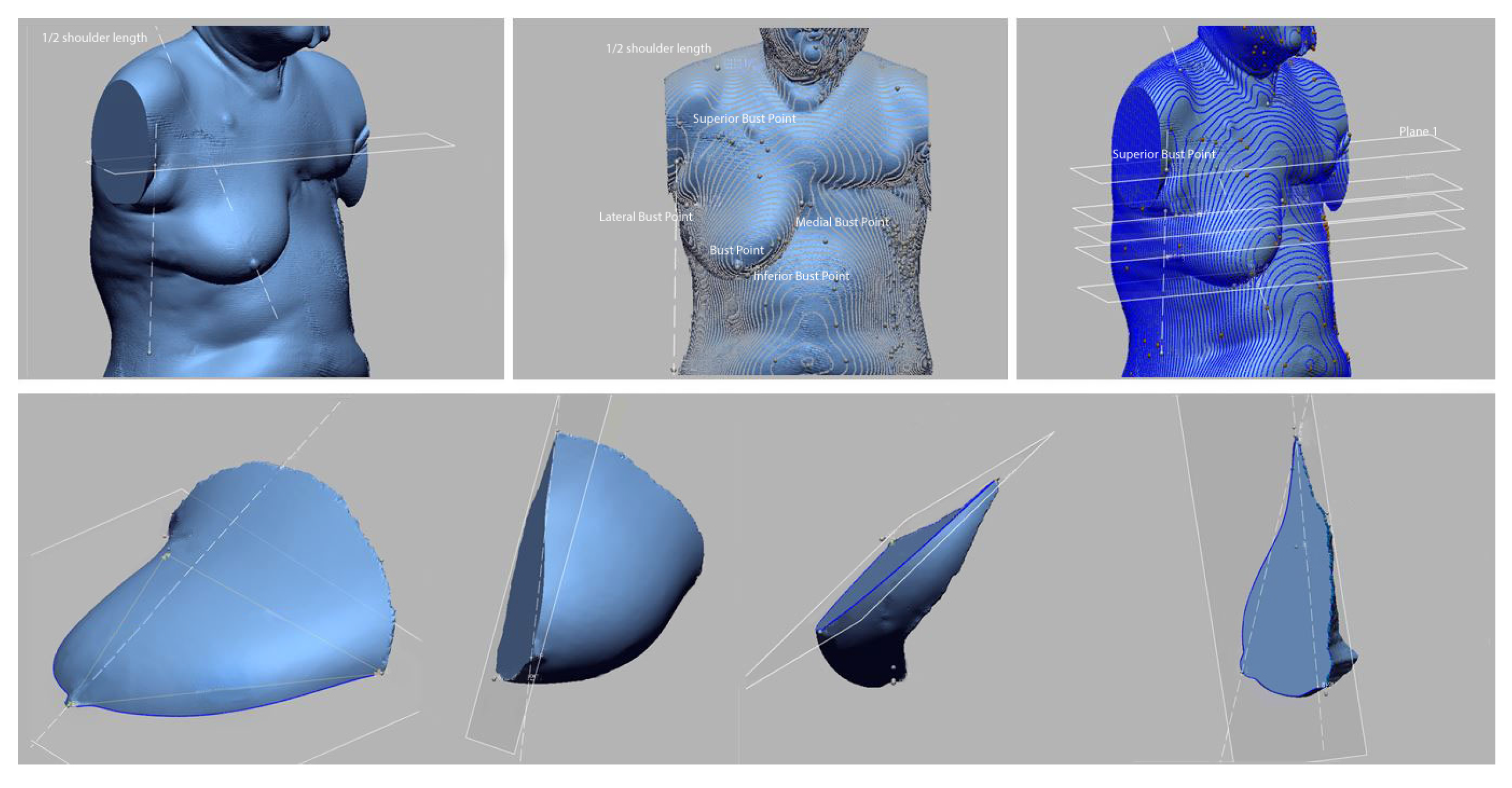
Learning the shape of female breasts: an open-access 3D statistical shape model of the female breast built from 110 breast scans

A combined PET-MRI scan could improve treatment for patients with early breast cancer
Frontiers Accelerated partial breast irradiation in early stage breast cancer
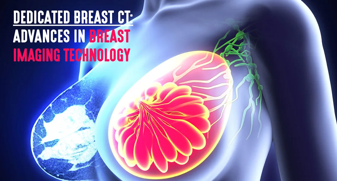
Dedicated CT Imaging of the Breast
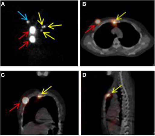
Frontiers Definition of Internal Mammary Node Target Volume Based on the Position of the Internal Mammary Sentinel Lymph Nodes Presented on SPECT/CT Fusion Images
Assessment of Three Breast Volume Measurement Techniques: Single Marking, MRI and Crisalix 3D Software®

Mamadou DIOP, Professor (Assistant), PhD Physics (Optics), The University of Western Ontario, London, UWO, Department of Medical Biophysics
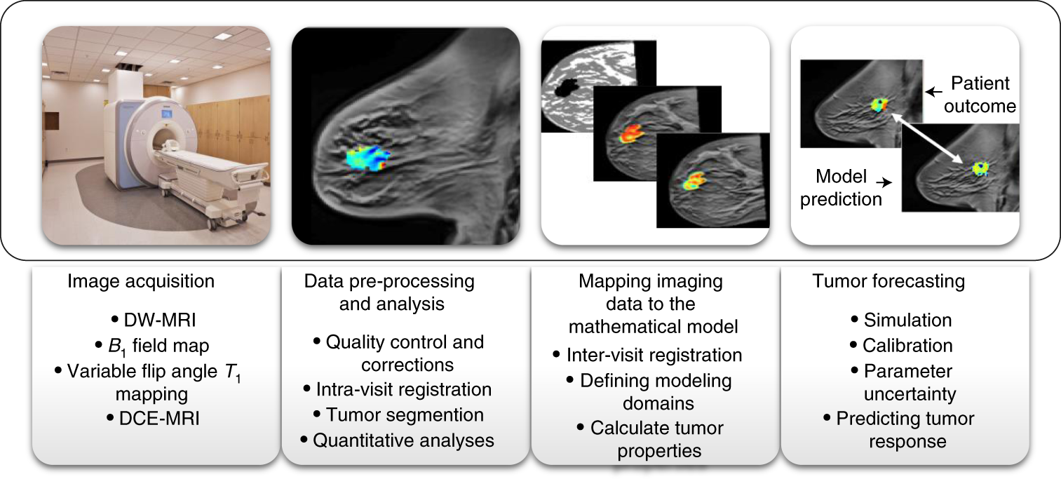
Quantitative magnetic resonance imaging and tumor forecasting of breast cancer patients in the community setting

PDF) Structured-light surface scanning system to evaluate breast morphology in standing and supine positions
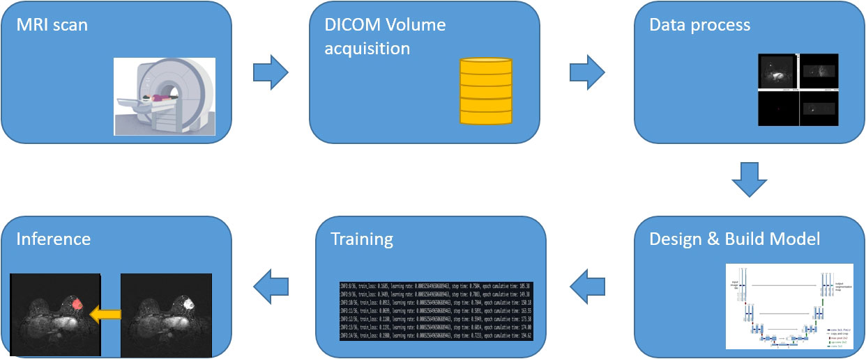
Frontiers Deep learning-based automatic segmentation for size and volumetric measurement of breast cancer on magnetic resonance imaging
Tomography, Free Full-Text

Abbreviated MRI shows promise, CEM has tradeoffs in breast imaging
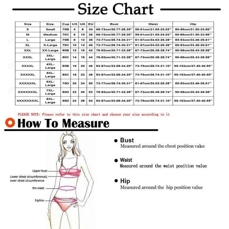
- KBKYBUYZ Women's French Hot Gathering Large Size Bra Set Ultra-thin Big Breasts Shows Small Collection
- Granny Body Women's Sports Bra Big Chest Small Running Shockproof Gathering No Steel Ring Sports Bra Plus Size Body Suit at Women's Clothing store

- Front Buckle Underwear No Steel Ring Anti-sagging Breastfeeding Large Size Thin Big Breasts Small Breasts Gathered Beautiful Back Bra
- CLZOUD Bra for Woman Big Size Lace Women Full Cup Thin Underwear
- Bras for Women Bra Women's French Elegant Gathering Large Size Bra Set Ultra thin Big Breasts Shows Small Collection Bras for Women Period Underwear New Arrival White,L